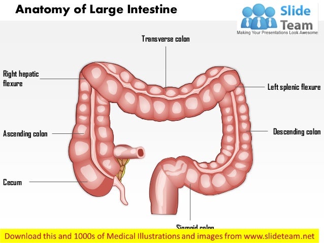
These sections form an arch, which encircles the small intestine. It receives digested food from the small intestine, from which it absorbs water and electrolytes to form faeces.Īnatomically, the colon can be divided into four parts - ascending, transverse, descending and sigmoid. The colon (large intestine) is the distal part of the gastrointestinal tract, extending from the cecum to the anal canal. This allows toxins absorbed from the colon to be processed by the liver for detoxification. The superior mesenteric and inferior mesenteric veins ultimately empty into the hepatic portal vein.

The neurovascular supply to the colon is closely linked to its embryological origin: The long length of the mesentery permits this part of the colon to be particularly mobile. The sigmoid colon is attached to the posterior pelvic wall by a mesentery – the sigmoid mesocolon. This journey gives the sigmoid colon its characteristic “S” shape. The 40cm long sigmoid colon is located in the left lower quadrant of the abdomen, extending from the left iliac fossa to the level of the S3 vertebra. When the colon begins to turn medially, it becomes the sigmoid colon. It is retroperitoneal in the majority of individuals, but is located anteriorly to the left kidney, passing over its lateral border. Descending ColonĪfter the left colic flexure, the colon moves inferiorly towards the pelvis – and is called the descending colon. Unlike the ascending and descending colon, the transverse colon is intraperitoneal and is enclosed by the transverse mesocolon.

The transverse colon is the least fixed part of the colon, and is variable in position (it can dip into the pelvis in tall, thin individuals). Here, the colon is attached to the diaphragm by the phrenicocolic ligament. This turn is known as the left colic flexure (or splenic flexure). The transverse colon extends from the right colic flexure to the spleen, where it turns another 90 degrees to point inferiorly. This turn is known as the right colic flexure (or hepatic flexure), and marks the start of the transverse colon. When it meets the right lobe of the liver, it turns 90 degrees to move horizontally. The colon begins as the ascending colon, a retroperitoneal structure which ascends superiorly from the cecum. The colon averages 150cm in length, and can be divided into four parts (proximal to distal): ascending, transverse, descending and sigmoid.


 0 kommentar(er)
